


Conization Electrode | NJ Medical Instruments
In stock
Conization Electrode | NJ Medical Instruments
In stock
Conization Electrode | NJ Medical Instruments
In stock
Conization Electrode | NJ Medical Instruments
In stock
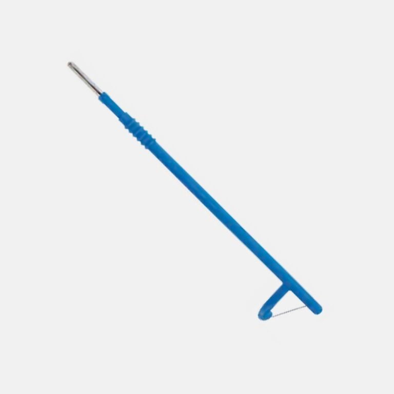
Conization Electrode Disposable | NJ Medical Instruments
In stock
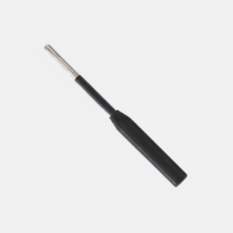
Extender Electrode | NJ Medical Instruments
In stock
Conization Electrodes: Taking a Cone-Shaped Cervical Specimen
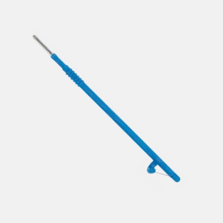
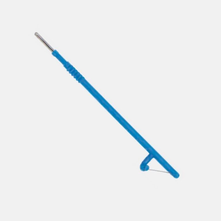
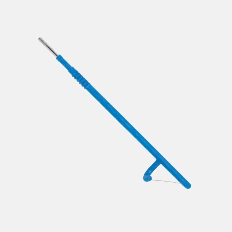
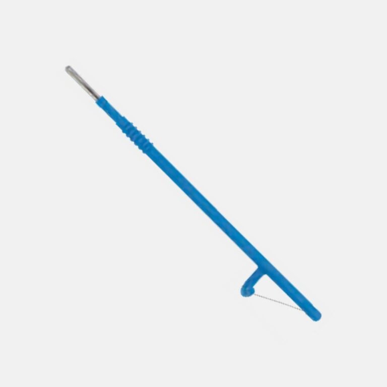
A conization electrode removes a cone of cervical tissue rather than a shallow disc, and that shape is the point. Abnormal cells do not always stay on the visible surface of the cervix — where they extend upward into the endocervical canal, a standard shallow loop excision removes the surface lesion and leaves disease behind at the top. A conization electrode is shaped so the excision narrows as it goes deeper, following the canal and taking the full height of the affected zone in one piece. Removing it intact matters because the pathologist assesses the margins: a specimen that comes out fragmented cannot be reported with the same confidence about whether the disease was fully excised. NJ Medical Instruments manufactures the range in surgical-grade stainless steel and tungsten wire, supplied worldwide direct from the factory for individual purchase and for bulk, wholesale, and OEM/private-label supply.
Cone geometry and depth
Electrodes are specified by the depth and base width of the cone they produce. A shallower cone suits a lesion largely confined to the ectocervix. A deeper cone is needed where the transformation zone lies further into the canal or is not fully visible on colposcopy. The clinical trade-off is real and belongs to the surgeon: excising too little risks leaving disease at the margin, excising more than necessary removes cervical tissue that has a role in subsequent pregnancy, so the electrode is selected to match the assessed extent rather than defaulted to.
Specimen orientation
Because margin status guides what happens next, the specimen must reach the laboratory oriented. Many teams place a marker suture at a known position before or immediately after excision, so the pathologist can report which margin is involved rather than simply that one is. The electrode design supports this by producing a single intact cone rather than several fragments.
After the excision
A ball electrode is rolled over the crater in coagulation mode to achieve haemostasis of the cervical bed. Insulated speculums and lateral wall retractors are used throughout, because monopolar current in a confined conductive space can arc to the vaginal wall — non-insulated instruments should not be substituted while current is in use.
Which electrode do you need?
| Electrode | Shape | Used for |
|---|---|---|
| Conization electrode, shallow | Wide base, low cone | Ectocervical lesions |
| Conization electrode, deep | Narrow base, tall cone | Lesions extending up the canal |
| Square electrode | Square wire | Larger or deeper block excision |
| Loop electrode | Thin wire loop | Standard LEEP / LLETZ excision |
| Ball electrode | Spherical tip | Coagulating the cervical bed after |
| Insulated speculum | Coated blades | Preventing current arcing |
Explore related electrosurgical instruments
- Electrosurgical Instruments for Gynecology
- Electrodes (2.4 mm) and Electrodes (4.0 mm)
- Monopolar Cables and Patient Return Plates
- All Electrosurgical Instruments
All manufactured to international standards, with reusable and single-use options and worldwide shipping.
Bulk, wholesale & OEM supply
As a direct manufacturer, NJ Medical Instruments supplies conization and loop electrodes in bulk and wholesale quantities to hospitals, colposcopy clinics, and distributors, and produces OEM / private-label electrodes in custom cone depths and widths. Contact us by email at info@njmedicalinstruments.com or by WhatsApp for volume pricing — no minimum-order barriers, no middlemen.
Frequently asked questions
What is a conization electrode used for?
It removes a cone-shaped specimen of cervical tissue, taking the full height of the affected transformation zone including tissue extending up into the endocervical canal, in one intact piece for pathological assessment.
What is the difference between a conization and a LEEP loop electrode?
A LEEP loop takes a relatively shallow disc of the transformation zone. A conization electrode produces a cone that narrows as it goes deeper, used when the lesion extends up the endocervical canal beyond the reach of a shallow loop.
What cone depth should be selected?
Depth is matched to the assessed extent of the lesion. Too shallow risks leaving disease at the margin; deeper than necessary removes cervical tissue with a role in future pregnancy. The electrode is chosen against colposcopic assessment rather than defaulted to.
Why must the specimen be removed intact?
The pathologist reports on the margins to determine whether the disease was fully excised. A fragmented specimen cannot be assessed with the same confidence, which is why the electrode is designed to produce a single intact cone.
Why are insulated speculums used with these electrodes?
Monopolar current in the confined, conductive space of the vaginal canal can arc to the vaginal wall. The insulation confines the current to the electrode tip, and non-insulated instruments should not be substituted while current is in use.
Do you supply conization electrodes in bulk or for OEM / private label?
Yes. As a direct manufacturer we supply conization and loop electrodes in bulk and wholesale quantities and produce OEM / private-label electrodes in custom cone depths and widths. Contact us by email or WhatsApp for volume pricing.